Thousands of lives are saved every year through screening that detects breast cancer before the patient has any symptoms. Doctors agree that those numbers could increase with more widespread screening, but the tests can be uncomfortable and expensive.
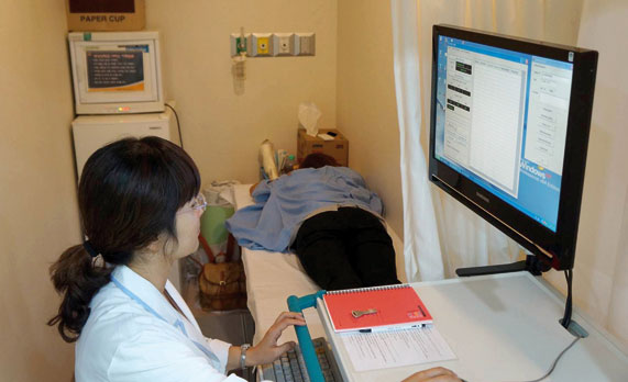
With microwave tomography imaging, the patient lies on a special table and places her breast in a compartment filled with gel. The technician then does the scan.
Two SDSU computer scientists—professor Sung Shin and assistant professor Wei Wang—have been collaborating with the Electronics and Telecommunications Research Institute, known as ETRI, in Daejeon, South Korea, to develop a more accurate, less expensive means of detecting breast tumors called microwave tomography imaging,
or MTI.
Microwave imaging may more accurately detect tumors, particularly in women with dense breast tissue, plus the imaging can be done for a fraction of the cost of current techniques, said Shin. Nearly 50 percent of U.S. women have dense breast tissue, making it harder to detect tumors using mammography, according to the American Cancer Society.
MTI offers an increased comfort level for the patient—unlike mammography, no compression techniques are necessary, Wang explained. Waves travel through a gel called Phantom, so the patient simply lies down on a special bed that allows her to place one breast at a time in the gel.
Greater accuracy, lower cost
Although mammography is the least costly evaluation technique, its accuracy suffers when dealing with dense breast tissues, Shin said. Another downside to mammography is the patient’s exposure to X-rays.
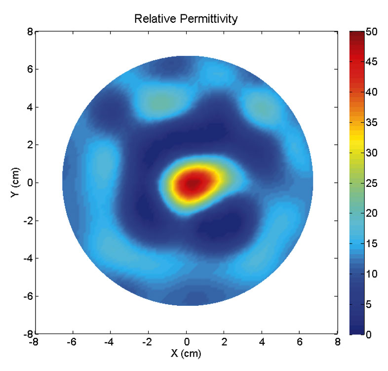
Software developed by SDSU computer scientists compares this microwave tomography image to an MRI of the same tumor and extracts the files of similar cases, which doctors use to determine the best treatment for a patient.
Tomography images are three-dimensional, so only one image is required, rather than the multiple angles necessary with mammography, Wang said. Microwave tomography can be a compromise between mammography and magnetic resonance imaging, commonly known as MRI.
The current cost of a tomography machine is less than $100,000, as compared to $300,000 for a mammography unit and anywhere from $2.5 to $4 million for an MRI machine, Shin said. However, what the experimental machine will eventually sell for has yet to be determined.
The government-owned South Korean research institute holds the patent on the microwave tomography machine. Researchers are developing its capabilities as a cancer screening tool and hope to market the equipment to U.S. health-care institutions.
This technology holds promise as a safe method for examining soft tissues, such as breasts, lungs, heart and brain. It may also be effective in detecting prostate cancer.
Designing software to compare images
The SDSU computer scientists have been developing software that first identifies the tumor on the microwave tomography image and then compares that to a database of more than 100,000 MRI images, Wang explained. Work on the five-year, $500,000 project began in 2010.
The program then chooses the cases that are most similar and extracts the image along with the case file. The cases selected tell doctors what treatments were used and how successful they were at combating the cancer. Based on the patient histories, doctors will have the information they need to determine the best plan of action.
The professors and their team of four master’s students and one undergraduate have developed several algorithms, Wang said, “to optimally identify the tumor.â€
The experimental imaging technique was approved for use on human subjects in Korea in 2011, Shin explained. On this portion of the international collaborative project, he is working with Dr. Wu-Kyung Moon of Seoul National University Hospital, one of South Korea’s leading cancer researchers.

Sung Shin
Wei Wang
In fall 2012, the first 15 patients were screened using MTI, MRI and mammography. This patient data allows the researchers to determine which computer program is most effective matching the MTI and MRI images.
Based on these comparisons, they will then improve the program’s capabilities by adjusting the algorithms.
Higher frequency, better images
The process, however, is dependent on the image quality that the machine produces, Shin explained. The imaging machine currently uses a wave frequency of three gigahertz. To improve the quality of the images, the research institute is upgrading its system to six gigahertz and will begin human testing in March.
“Higher frequency will give a better image,†said Wang. Even at the higher frequency level, the imaging process exposes patients to less radiation than a cell phone, an important safety consideration. The researchers hope that future funding will involve partnering with an American health-care facility.
Once the software and imaging technique are perfected, Shin and Wang believe that microwave tomography can improve the accuracy and reduce the costs of breast cancer screening.
Christie Delfanian


